Prevalence of Dens Invaginatus and Its Association with Periapical Lesions: A Study Using Cone‐Beam Computed Tomography
DOI:
https://doi.org/10.5281/zenodo.13623834Keywords:
Dens Invaginatus, Periapical Lesions, Cone Beam Computed TomographyAbstract
Objectives: This study aimed to determine the prevalence of Dens Invaginatus (DI) and examine the association between factors such as age, gender, and DI type with the occurrence of periapical lesions.
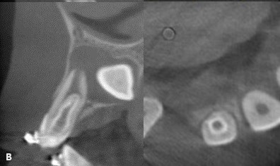
Materials and Methods: Cone beam computed tomography (CBCT) images of 250 patients were evaluated. The relationship between gender, tooth number, DI type according to Oehler classification, and periapical lesions (PL) was examined. PL were grouped using Estrela's CBCT Periapical Index (CBCT PAI). Periapical lesion incidence rates were statistically compared using chi-square tests and descriptive statistics. The statistical analysis was performed using SPSS v27.0 software.
Results: 250 CBCT volumes were examined. The study involved 32 patients (average age 29.15 ± 11.50 years). DI was found in 10.0% of maxillary lateral teeth (11.6% right, 8.4% left) with no significant gender or age group differences (p>0.05). Type I DI was most common in the 15-20 age group. Most teeth had a CBCT PAI=0, with no significant differences across age groups (p>0.05). However, for the right lateral tooth, Type II DI was significantly associated with higher CBCT PAI scores (p<0.05), whereas no significant difference was found for the left lateral tooth (p=0.142).
Conclusion: DI is a developmental dental anomaly that is relatively prevalent. The presence of associated periapical lesions and the proportion of maxillary lateral teeth impacted by DI should be meticulously assessed during the diagnosis and treatment planning process. Clinicians can more effectively plan and execute treatment by comprehending the prevalence of DI, its subtypes, and their relationship to periapical lesions.
Downloads
Published
How to Cite
Issue
Section
License
Copyright (c) 2024 Ayse Hanne Sarı, Güldane Mağat

This work is licensed under a Creative Commons Attribution-NonCommercial 4.0 International License.
CC Attribution-NonCommercial 4.0