
Endodontic management of a maxillary first molar with seven root canals using a dental operating microscope: A case report
DOI:
https://doi.org/10.5281/zenodo.14948031%20Keywords:
additional canals, dental operating microscope, upper first molar, root canal morphology, seven root canalsAbstract
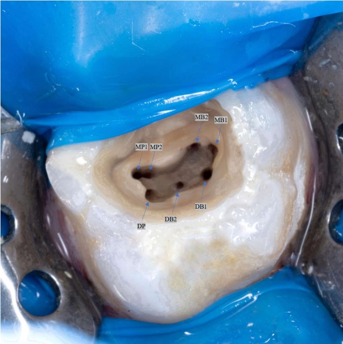
The main objective of root canal treatment is the thorough cleaning and shaping of the entire root canal system. For this, the detection of all canals is of significant clinical importance. Maxillary first molars (Mx1M) commonly present with three roots and three canals, with a second mesiobuccal canal (MB2) also present. With the advent of improved diagnostic systems, it has been reported that Mx1M can have different anatomical variations. This varying number of configurations presents a challenge to the endodontist in detecting and treating through the root canal. This problem can be avoided by using magnification systems or advanced diagnostic techniques. Although four canals were detected at the beginning of the treatment, a rare morphology and three additional canals were detected with the contribution of a dental operating microscope (DOM). This case report presents the endodontic management of an Mx1M with pulpal necrosis and symptomatic apical periodontitis. Nonsurgical endodontic therapy of a left Mx1M with three roots and seven root canals was successfully performed under a DOM.
Downloads
Published
How to Cite
Issue
Section
License
Copyright (c) 2025 ÖZGE BAŞAR

This work is licensed under a Creative Commons Attribution-NonCommercial 4.0 International License.
CC Attribution-NonCommercial 4.0