Evaluation of Maxillary First Molar Teeth's Mesiobuccal Root and Root Canal Morphology using two classification systems amongst a Turkish population: A Cone-beam Computed Tomography study
DOI:
https://doi.org/10.5281/zenodo.8306179Keywords:
cone-beam computed tomography, endodontics, tooth root, molar, classificationAbstract
Objectives: This research aimed to analyze and compare the morphology of the mesiobuccal (MB) root and its canals in maxillary first molars (M1Ms) using Vertucci (1984) and Ahmed et al. (2017) classification systems.
Materials and Methods: 250 cone beam computed tomography (CBCT) images of 500 M1M teeth were evaluated for MB root and canal configurations. The images were analyzed from sagittal, axial and coronal perspectives. Canal number and morphology were documented according to Vertucci's method as well as a more recent classification system.
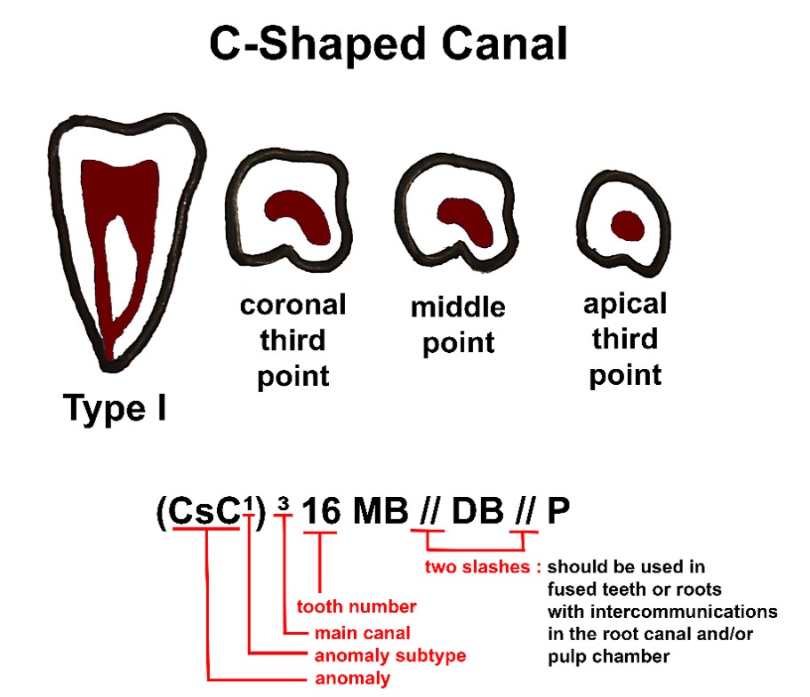
Results: The majority of MB roots had Type I morphology according to the Vertucci classification (right: 38.4%; left: 43.2%), and according to the new root canal morphology classification system, the most common code detected was 316 MB1 in the right side (37.6%), 326 MB1 for the left side (41.2%). Subsequently, Type IV (right: 24.4%; left: 26.0%) and Type V (right: 16.4%; left: 14.4%) were the next most frequently identified morphologies according to the Vertucci classification, whereas according to Ahmed's classification 316 MB2 (24.4%), 326 MB2 (25.2%), 316 MB1-2 (16.4%), and 326 MB1-2 (14.0%) were the most common.
Conclusion: It is vital for dentists to locate and treat all parts of a tooth, especially the MB2 canals in M1Ms, to prevent endodontic treatment failure due to microbial contamination and infection. For clinicians seeking clarity in root and canal morphology, the new classification system offers a more precise and user-friendly approach than the traditional Vertucci classification.
Downloads
Published
How to Cite
Issue
Section
License
Copyright (c) 2023 Güldane Mağat, Sultan Uzun, Glynn Dale Buchanan

This work is licensed under a Creative Commons Attribution-NonCommercial 4.0 International License.
CC Attribution-NonCommercial 4.0